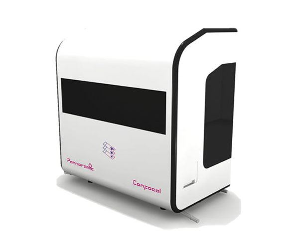
Your immunofluorescent samples will appear on your screen in unprecedented quality 3DHISTECH presents its latest addition to research pathology. By combining confocal imaging with award-winning whole slide scanning technology, your immuno-fluorescent samples will appear on your screen in unprecedented quality!
With 3DView, the digital 3D reconstruction of the fluorescent images gives an amazing insight view of the whole specimen.
Features
Innovative structured illumination confocal imaging to overcome the limitations of spinning pinhole-disc techniques. This delivers the highest light efficiency with minimal bleaching and fastest scanning speed.
Colocalized Fluorescent and Brightfield imaging
Automated immersion for high NA water immersion objective
Brightfield / Darkfield / Fluorescent Preview
Motorized Objective changer
1D and 2D barcode reading
SW DDIC (Digital Differential Interference Contrast) for low contrast brightfield visualization
Advanced export options (ROI, Wholeslide, grayscale/color, multichannel)
Specifications
Slide capacity 12 slides
Acceptable slide formats 25.5 (+-0.5) mm x 75.5 (+-0,5) mm, 1(+-0.05) mm thickness
Default objectives Zeiss Plan-Apochromat 20x/0.8 NA,
Zeiss C-Apochromat (W) 40x/1.2 NA
Camera type 5.5Mpx, 16 bit, low noise (1.3 e-)
PCO edge cooled scientific CMOS camera
Image resolution (in focus plane) 0,4 µm FWHM (with 40x 1.2NA objective)
Confocal sectioning 1,43 µm FWHM (with 40x 1.2NA objective)
Fluorescent illumination 6 channels Solid state light engine,
15000 hrs lifetime
Default fluorescent filter sets
# filter cube positions
filter type
Quad band: DAPI/FITC/TRITC/Cy5,
3 (BF+FL mode) or 4 (FL mode)
single-/dual-/quad-band
Brightfield illumination 3CCD equivalent separated R-G-B LED
Digital slide format Proprietary digital slide format (MRXS) with lossless or JPG/JPGXR/JPG2000 encoding
Export opinions single/multi chanel
area
file format
annotation or whole slide
TIFF/JPEG
Instrument dimensions
W x D x H 97 cm x 58 cm x 103 cm or 39"x23"x41"
Weight 100 kg

