
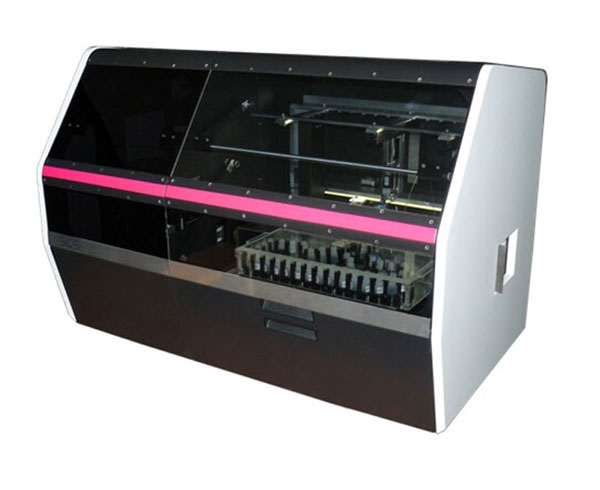
Staining - Coverslipping - Scanning unit: 3DHISTECH is the first company to combine 3 very important steps of the pathology workflow in one single device!
3DHISTECH introduces its revolutionary solution for integrated histo-pathology slide staining and digitalization (scanning) by a single platform. It does save valuable laboratory space and time and a top of that - thanks to its unique features - it goes significantly reduces reagent costs in order to keep laboratory operating expenses low. The patented automatic sample detection makes only the effective sample area get stained exatly the desirable level of reagent and thousand of serial staining become the identical intensity.
Efficiency
Lower laboratory operational expenses, reduces reagent and antibody consumption, operational expense saving by 50% accelerated testing, laboratory space saving.
Simplicity
Full automation, overall workflow control, LIS connectivity, easy handling of consumables. Remote management and maintenance, integrated design, digital analysis and immediate automated test reporting.
High quality
Very high resolution digital slide scanning, no cross-contamination of samples and reagents, no dilution, in-built waste management, thousands of serial stainings with identical intensity, only the detected sample area get stained using exactly the esired level of reagent.
Flexibility
IHC and FISH, fluorescent features, digital slide preview, 72 slide capacity and 40 different reagent and antibody capacity, open system: your own primary antibody can be used, modulare construction, parallel task execution.
Features
• single device integrating an immunostainer,
• a coverslipper and a scanner
• the patented automatic sample detection makes
• only the effective sample area gets stained
• fully automated workflow from unstained tissue
• to the quantitative IHC evaluation on digital slide
• first stained slides in CaseCenter can be available
• on-line right away: speedy evaluation
• iSaCS scanning device can be used separatly as
• individual scanner
• Image analysis process will be run automatically on
• the whole slide
• Slide and protocol recognition based on bar code
• Standardization of IHC/IF stainings
Technical Specifications
Slide capacity 72/36 slides/5 hours
Max. number of reagents 40
Coverslipper 150 pcs glass coverslips
22 x 50 mm
Reagent identification 2D barcode
Acceptable slide formats 25 x 75 mm,
1 mm thickness
Brightfield and fluorescent scanning Yes/Yes
Available magnifications 25x, 40x
Fluorescent illumination 6 channels, 15,000 hrs lifetime
Digital slide format Proprietary digital slide format
(MRXS) with JPG, JPG2000 encoding
Power requirements 100-240 VAC, 350W
W x D x H (cm) 112 x 66 x 78
Weight (kg) 120

Integrated Immunohistochemistry Workflow
Integrated Immunohistochemistry Workflow optimizes reagents and antibody resources, minimizes the turn over period and maximizes the quality and reliability.
Digital pathology enables staining integrated with automated evaluation and analysis.
The main challenge is to automate the routine detection of tissue antigen markers by using the advantage of digital imaging technology in the immunohistochemical working flow.
iSaCS is combining the fully automated unique digital microstaining with the world-class digital slide scanning and software base evaluation technique.
iSaCS user interface
The patented automatic sample detection makes only the identified sample are get stained exactly the desirable level of reagent. This way it significantly reduces reagent costs in order to keep the laboratory operating expenses low.